

FEI Co.
Hillsboro, OR 97124

STEM System analyzes multiple wafer samples.
Designed for semiconductor labs, Expida(TM) 1255S DualBeam(TM) System integrates STEM Scanning/Transmission Electron Microscopy sample preparation with high-resolution imaging and analysis in single tool. It features ion beam column for preparing TEM samples, and electron column with 14-segment STEM detector for 30 kV imaging. System assures correct end-pointing and precise lamella thickness by...
Read More »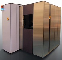
Wafer Handling System offers sub-45 nm process control.
Expida(TM) 1255S wafer DualBeam(TM) system integrates wafer-level STEM (scanning/transmission electron microscopy) sample preparation with imaging and analysis capabilities. Equipped with ion beam column for preparing TEM samples and electron column with 14-segment STEM detector for high-resolution 30 kV imaging, system ensures correct end-pointing and precise lamella thickness by enabling STEM...
Read More »FEI Expands Helios Nanolab(TM) Family for Semi Market
NanoLab 400/400S to be Introduced at SEMICON Japan Giving Users a Complete Range of Advanced Solutions for Semiconductor Labs NOVEMBER 30, 2006/Hillsboro, Ore.-FEI Company (Nasdaq: FEIC) will expand its top-of-the-line Helios NanoLab(TM) family of DualBeams(TM) when it introduces the Helios NanoLab 400 and 400S systems next week at SEMICON Japan. Combining advanced focused ion beam (FIB) and...
Read More »FEI Receives $11.5 Million Order from Technical University of Denmark
Seven Systems to Form the Core of DTU's New Center for Electron Nanoscopy and Pave the Way for Advanced Catalyst Research NOVEMBER 27, 2006/Hillsboro, Ore.-The Technical University of Denmark (DTU) has placed a $11.5 million dollar order for seven FEI microscopes that will form the core of the University's new Center for Electron Nanoscopy (CEN). The order represents the largest product sale ever...
Read More »FIB/SEM Systems are designed for semiconductor labs.
NanoLab(TM) 400/400S feature high resolution field emission scanning electron microscope column combined with Sidewinder(TM) focused ion beam column and gas chemistries. Suited for sub-65 nm node devices, DualBeams(TM) systems offer low-kV SEM resolution to support 3D cross-sectional imaging and analysis. NanoLab 400 comes with high resolution stage/load lock, while Model 400S is equipped with...
Read More »Imperial College London Unveils UK's First Titan (TM) S/TEM
World's Most Powerful Commercially-Available Microscope Provides Access to Atomic-Scale Data for Nanotechnology Research OCTOBER 18, 2006/Hillsboro, Ore.--Imperial College London has unveiled one of the UK's most powerful microscopes, the Titan(TM) 80-300 S/TEM from FEI. It is the world's most powerful commercially-available scanning/transmission electron microscopes and one of the only...
Read More »FEI Company and Malvern Instruments Team
FEI's Quanta(TM) SEMs and Malvern's Advanced Particle Analysis Software Combine to Deliver Ground-breaking Solution for Nanoscale Applications Hillsboro, Ore./September 12, 2006 --- FEI Company (NASDAQ: FEIC) and Malvern Instruments Ltd (Malvern, UK) have entered into a joint development and marketing program for advanced nanoparticle analysis utilizing Malvern's particle image analysis software...
Read More »FEI Receives Titan(TM) Order from Japanese Steelmaker
Steel Researchers to Utilize World's Most Powerful Microscope for Advanced Materials Characterization and Analysis HILLSBORO, Oregon/September 4, 2006-FEI (Nasdaq: FEIC) today announced that Japan's JFE Steel Corporation has ordered a Titan(TM) 80-300 for its research center in Kawasaki. JFE Steel, a leading global supplier of steel products, is the first Japanese customer to order the Titan...
Read More »FIB/SEM System offers nanoscale imaging and analysis.
Helios NanoLab(TM) DualBeam(TM) features ultra-high resolution field emission scanning electron microscope (SEM) column combined with Sidewinder(TM) focused ion beam (FIB) column and gas chemistries to provide imaging resolution and contrast in DualBeam system. Small DualBeam platform enables 3D characterization, analysis, and image reconstruction applications, nano-prototyping (fabrication and...
Read More »FEI Launches Certified Tools Program Globally
New Program Offers Customers Enhanced Flexibility for Purchasing Factory Certified FEI Solutions HILLSBORO, Ore., July 10 / -- FEI Company (NASDAQ:FEIC) has announced the global launch of its Certified Tools program featuring factory-refurbished FEI systems that are fully-tested and warranted to meet original factory specifications. With the Certified Tools program, customers will now have...
Read More »