X-ray CT Scanner obtains video images of living organs.
Share:
Press Release Summary:
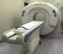 Equipped with 256-row detectors, 4-dimensional X-ray computed tomography (CT) scanner provides accurate images of organs. It can measure locations of moving organs, such as lungs or liver, in real-time by increasing detector revolving speed. Able to image entire heart or brain, product is suited for use in such applications as diagnosis of heart diseases and heavy particle radiotherapy for lung cancer.
Original Press Release:
National Institute of Radiological Sciences, Toshiba Medical Systems Jointly Develop Four-dimensional X-ray CT Scanner Enabling to Obtain Video Images of Living Organs
Tokyo (JCNN) - The National Institute of Radiological Sciences (NIRS) announced on July 7 that the NIRS, in collaboration with Toshiba Medical Systems, has developed the world's first four-dimensional X-ray computed tomography (CT) scanner to provide accurate images of organs and unveiled images created by the new device.
The new scanner, which has 256-row detectors, can image the entire heart or brain. In addition, by increasing the revolving speed of detectors, the scanner can measure the locations of moving organs such as lungs or liver in real time.
With a view to applications in the diagnosis of heart diseases and heavy particle radiotherapy for lung cancer, the NIRS has commenced clinical research using the scanner. The NIRS expects that the new scanner will contribute to the development of sports medicine.