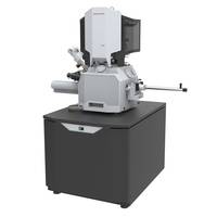
Aquilos FIB/SEM System is used for tomographic imaging.
Press Release Summary:

Aquilos FIB/SEM cryo-DualBeam System is used for preparation of frozen, thin lamella samples from biological specimens. Unit consists of focused ion beam and scanning electron microscope. Product enables user to create the samples with precisely-controlled thickness. System’s Electron tomography computes a three-dimensional model of a single molecular structure and provides near-atomic scale resolution.
Original Press Release:
Industry’s First Dedicated Cryo-DualBeam System Automates Preparation of Frozen, Biological Samples
New Thermo Scientific Aquilos FIB/SEM protects sample integrity and enhances productivity for cryo-electron tomography workflow
The new Thermo Scientific Aquilos is the first commercial cryo-DualBeam (focused ion beam/scanning electron microscope) system dedicated to the preparation of frozen, thin lamella samples from biological specimens for high-resolution tomographic imaging in a cryo-transmission electron microscope (cryo-TEM).
“The Aquilos completes our cryo-electron tomography workflow, allowing customers to reliably create the samples with precisely-controlled thickness with minimal investment in time and effort,” said Peter Fruhstorfer, vice president and general manager, life sciences, Thermo Fisher Scientific. “The ability to properly prepare the thin lamella samples, while at the same time maintaining the necessary temperature, vacuum and transfer conditions, has been a significant challenge for researchers. With the Aquilos, we have essentially removed that impediment, making tomography accessible to a much broader community of researchers and scientists.”
Elizabeth Villa, assistant professor of biophysics, University of California San Diego, has been using the Aquilos cryo-DualBeam in her work on macromolecular complexes, “Cryo-electron tomography’s ability to visualize structures in their native context allows researchers to observe functional relationships and interactions with other components in the cellular environment,” said Dr. Villa. “This technique promises to become an important tool for scientists seeking a better understanding of living systems at the molecular level.”
Electron tomography computes a three-dimensional (3D) model of a single molecular structure in its native, fully functional context by combining multiple images from different perspectives, much like a medical X-ray CT (computed tomography) scan. It is a powerful complement to single particle analysis (SPA), which can provide near-atomic scale resolution but requires a large collection of isolated identical particles. Together, these techniques can provide biologists with a more complete picture of a protein’s structure and function.
About Thermo Fisher Scientific
Thermo Fisher Scientific Inc. is the world leader in serving science, with revenues of $18 billion and more than 55,000 employees globally. Our mission is to enable our customers to make the world healthier, cleaner and safer. We help our customers accelerate life sciences research, solve complex analytical challenges, improve patient diagnostics and increase laboratory productivity. Through our premier brands - Thermo Scientific, Applied Biosystems, Invitrogen, Fisher Scientific and Unity Lab Services - we offer an unmatched combination of innovative technologies, purchasing convenience and comprehensive support. For more information, please visit www.thermofisher.com.
Contact:
Chuck Anderson
Thermo Fisher Scientific
+1 781-622-1348


