Objective Lens suits deep imaging of thick biological samples.
Share:
Press Release Summary:
Featuring working distance of 4 mm, XLPLN25XSVMP SCALEVIEW 25x Objective Lens is optimised to match refractive index properties of SCALEVIEW-A2 clearing agent, which renders samples nearly translucent while preserving fluorescent signals, to produce maximum image quality, sharpness, and brightness. SCALEVIEW integrates with Olympus's FluoView FV1000MPE multiphoton microscopes and FV10-ASW software v3.1, providing power to visualize 3D structures in morphologically intact tissue.
Original Press Release:
The New Olympus XLPLN25XSVMP SCALEVIEW Objective Lens with 4mm Working Distance for Deep Biological Imaging
Look deep into your samples, without the need for sectioning
Hamburg, - Olympus has today released the new XLPLN25XSVMP SCALEVIEW 25x objective lens (NA 1.0) for the deep imaging of thick biological samples. The revolutionary SCALEVIEW approach was developed in collaboration with the RIKEN Brain Institute in Japan, where it allowed researchers to create highly accurate 3D structural representations of brain tissue. Until now, the use of such samples has been limited by the effects of tissue opacity and light scattering. To counter this, the new objective has a super-long-working-distance of 4mm and works alongside Olympus's new SCALEVEW-A2 clearing agent, which renders samples nearly translucent while preserving fluorescent signals. Image quality, sharpness and brightness are then maximised as the objective is optimised to match the refractive index properties of the clearing agent. When used as part of a complete Olympus FluoView FV1000MPE multiphoton system, the new objective makes it possible to peer deeper into samples than ever before, allowing you to generate truly insightful results from intact specimens.
The SCALEVIEW 25x objective and SCALEVIEW-A2 reagent are designed to boost the capabilities of multiphoton microscopy, allowing accurate reconstructions up to a depth of 4mm to be generated using fluorescent markers. Previously, the investigation of thick or complex samples such as brain tissue had required that analysis be carried out using thin tissue sections. Mechanical slicing of tissue into very thin sections requires elaborate protocols and challenging data reconstruction methods to visualize exactly how the slices fit together. By clearing formalin-fixed tissues and allowing light to pass through the sample, the SCALEVIEW-A2 reagent minimises the need for sectioning, allowing the user to generate insightful data that more accurately reflects the true internal structure of a complex specimen. Using the technique, true 3D representations can be created, without the need for complex interpolation, predictive algorithms or guesswork.
To maximise the advantages provided by the SCALEVIEW-A2 clearing solution, Olympus has released the SCALEVIEW 25x objective, which has been specifically designed for deep imaging. This dedicated multiphoton objective with an ultra-long working distance enables the high-precision imaging of transparent biological specimens to a depth of 4mm. The objective is equipped with a correction collar and is optimised to work best with the SCALEVIEW-A2 reagent (refractive index 1.38), facilitating the production of detailed, crisp images. As part of a complete system, the new SCALEVIEW approach integrates seamlessly with Olympus's FluoView FV1000MPE multiphoton microscopes and the FV10-ASW software v3.1, providing the power to visualise 3-dimensional structures at unprecedented depths in morphologically intact tissue.
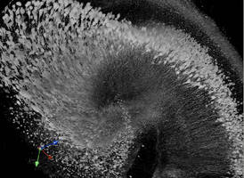
The image shows a 3D representation of YFP-expressing neurons in an excised mouse hippocampus, created using images captured with an Olympus FluoView FV1000MPE multiphoton microscope system using an XLPLN25XSVMP SCALEVIEW objective lens (image provided courtesy of Hiroshi Hama and colleagues at the RIKEN Brain Science Institute, Japan).