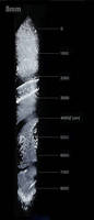
Objective Lens images 8 mm deep into biological samples.
Share:
Press Release Summary:
With 8 mm working distance, 0.9 NA, and correction collar, XLSLPLN25XSVMP SCALEVIEW 25x objective lens enables deep imaging of tissues without requiring potentially damaging micro-dissections. Product works with SCALEVIEW-A2 clearing reagent and enables crisp, clear imaging of structures, such as neurons. When used with FluoView FV1000MPE multiphoton microscope, lens lets users obtain complete system that avoids occurrence of artifacts due to slicing.
Original Press Release:
The New Olympus SCALEVIEW Super-Long-Working-Distance Objective Images 8 mm Deep into Biological Samples
Looking deeper into intact samples
Hamburg - Olympus has introduced the new XLSLPLN25XSVMP SCALEVIEW 25x objective lens with an 8 mm working distance, enabling the deep imaging of tissues, without the need for potentially damaging micro-dissections. Designed for use with the SCALEVIEW-A2 clearing reagent, this new objective lens enables crisp and clear imaging of structures, such as neurons extending 8 mm deep into tissues, much further than possible before. Optimised for use with the Olympus FluoView FV1000MPE multiphoton microscope, users can obtain a complete system which avoids the occurrence of artefacts due to slicing, thus enabling you to be confident in the biological relevance of your findings.
Following the recent introduction of the SCALEVIEW 4 mm objective lens, the 8 mm objective, with an NA of 0.9, enables users to look deep into samples. Both objectives are equipped with a correction collar and have been optimised for use with the SCALEVIEW-A2 reagent (refractive index 1.38), which essentially clears bodily tissues of their opacity to provide a clearly defined image. By making the tissue transparent, while simultaneously minimizing light scattering, images can be obtained from 8 mm below the surface of the sample. Through the elimination of tissue slicing, it is much easier for researchers to visualise how neural filaments connect in the brain for example; a process which is quite difficult when thin sections need to be pieced together to provide an accurate representation. As such, the SCALEVIEW objective lenses, in combination with the SCALEVIEW-A2 reagent and FluoView FV1000MPE microscope, provide a perfect option for developmental biology studies, as well as imaging and mapping of the brain and other organs.
The revolutionary SCALEVIEW approach was developed in collaboration with the RIKEN Brain Institute in Japan, where it allowed researchers to create highly accurate 3D structural representations of brain tissue. As part of a complete system, the new SCALEVIEW approach integrates seamlessly with Olympus's FluoView FV1000MPE multiphoton microscopes and the FV10-ASW software v3.1, providing the power to visualise 3-dimensional structures at unprecedented depths in morphologically intact tissue.