Multiphoton Confocal Microscope integrates 2 NLO lasers, OPO.
Share:
Press Release Summary:
Multiphoton confocal microscopes permit simultaneous use of 2 NLO lasers or one laser with optical parametric oscillator (OPO). In dual laser systems, different laser wavelengths simultaneously excite fluorescent dyes or proteins. Users can image specimens with one wavelength and manipulate them in multiphoton mode with another. OPO increases excitation range to up to 1,300 nm, covering absorption peak of red fluorescent proteins and enabling specimen protection.
Original Press Release:
New Possibilities for Multiphoton Microscopy from Carl Zeiss
OPO and simultaneous lasers for LSM 7 series
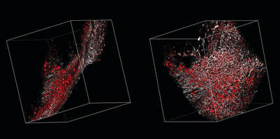
Jena, Germany:: Multiphoton confocal microscopes from Carl Zeiss now permit the simultaneous use of two NLO lasers or one laser with an optical parametric oscillator (OPO). Both components are fully integrated and expand the functionality of the multiphoton systems.
In dual laser systems different laser wavelengths simultaneously excite several fluorescent dyes or proteins. Without time loss, users can image specimens with one wavelength and manipulate them in the multiphoton mode with another. The automatic free beam adjustment gives the system a high degree of stability and reproducibility and ensures exact overlay of the two excitation beams. Such dual laser systems are used, above all, in intravital microscopy, e.g. for examining functional correlations in the brain of a mouse.
An OPO increases the excitation range of multiphoton microscopy to up to 1300 nanometers and therefore covers in particular the absorption peak of red fluorescent proteins such as mCherry, mPlum and tdTomato. This efficient, long-wave excitation enables excellent specimen protection. The potentially very high light intensities of the OPO lasers interact with specific structures in the tissue, leading to a doubling and tripling of the oscillation frequency. This non-linear effect of frequency doubling (SHG) occurs, for example. in striated skeletal muscle and collagen. Frequency tripling is especially visible on regions where structures with inconsistent optical density converge. These include lipid-water boundaries - for example, between membrane and cytoplasm.
About Carl Zeiss
Carl Zeiss Microscopy, LLC, offers microscopy solutions and systems for research, routine, and industrial applications. In addition, Carl Zeiss Microscopy markets microscopy systems for the clinical market, as well as optical sensor systems for industrial and pharmaceutical applications. Since 1846, Carl Zeiss has remained committed to enabling science and technology to go beyond what man can see. Today, Carl Zeiss is a global leader in the optical and opto-electronic industries.
With 12,872 current employees and offices in over 30 countries, Carl Zeiss is represented in more than 100 countries with production centers in Europe, North America, Central America and Asia. For more information on the breadth of solutions offered by Carl Zeiss Microscopy, please visit http://www.zeiss.com/micro.