Microscope is designed for electrophysiological experiments.
Press Release Summary:
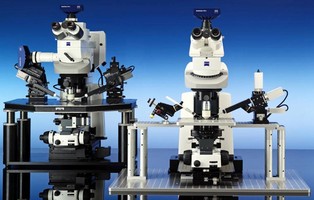
Optimized for flexibility and safety, Axio Examiner fixed stage microscope supports configuration with 4 upper parts, 2 lower parts, as well as various components - stages, micropipettes, light sources - and motorization options. Several interfaces are also available, and specimen area is extendable to free working distance of up to 100 mm. Connection of up to 2 AxioCam cameras and use of AxioVision v4.7 software with physiology module facilitate quantitative evaluation.
Original Press Release:
Carl Zeiss MicroImaging, Inc. Announces the Axio Examiner, Tailor-made for Electrophysiological Experiments
Thornwood, NY - Electrophysiological experiments can be performed with the Axio Examiner Fixed Stage Microscope from Carl Zeiss with considerably greater ease and with maximum flexibility and safety.
In the field of the neurosciences, Axio Examiner is particularly well suited for patch clamp experiments on nerve cells, examinations of brain sections and for measuring electrical signals on cells. Together with the new ZEISS LSM 710 NLO Laser Scanning Microscope, it is integrated into a multiphoton system with unmatched sensitivity. The connection of one or two AxioCam camera(s) and the use of the AxioVision 4.7 software with a special physiology module make the quantitative evaluation of typical experiments very comfortable and convenient. This includes the ability to visualize IR DIC or IR Dodt and Fluorescence in individual and merged live windows.
Axio Examiner has been designed so that complex experimental setups, e.g. with various stages, micropipettes or light sources are easy to set up, adapt and safe to use. This is ensured by the extremely stable stand design and by the slim profile in the front area of the system, providing the user with optimum access to the specimens. Axio Examiner offers outstanding flexibility in adapting the specimen area and in the selection of the contrasting method.
For configuring his or her specific Axio Examiner, the user has four upper parts, two lower parts and a large number of different components and motorization options to choose from. Numerous interfaces allow flexible adaptation to the respective requirements. The objective changer can be just as easily removed as the stage carrier and the condenser carrier. The specimen area is flexible and, depending on the system configuration, is extendable to a free working distance of up to 100 millimeters so that electrophysiological processes can be conveniently examined not only on living tissue sections and organs, but also on whole organisms. Settings can be changed without difficulty during the experiment as all relevant controls are arranged at the front of the system.
The optical design developed for Axio Examiner also offers maximum optical quality for transmitted light techniques and for advanced fluorescence applications. With the W N-ACHROPLAN and W Plan-APOCHROMAT series, water immersion objectives specially developed to meet the requirements of neuroscience are available for visible light and infrared. Various contrasting methods such as Dodt gradient contrast are options which are integrated in the stand design. Depending on the specimen, this makes it possible to achieve the highest resolution, the best contrast and optimum structure recognition in deeper tissue layers.
In addition, several motorized functions are available and can be remote-controlled via the docking station or the AxioVision software. All motors are automatically deactivated after the target position has been reached. In addition, the motors can be actively grounded. This guarantees that any remaining voltage can also drain off. The new Aquastop for condensers effectively protects the system against overflowing liquids. Axio Examiner offers flexible possibilities for additional magnification, e.g. with the double camera tube featuring an integrated continuous zoom system or a special additional magnification turret with fixed magnification steps.
About Carl Zeiss
Carl Zeiss MicroImaging, Inc., a subsidiary of Carl Zeiss, Inc., offers microscopy solutions and systems for research, laboratories, routine and industrial applications. In addition, Carl Zeiss MicroImaging markets microscopy and digital pathology systems for the clinical market, as well as spectral sensors for industrial and pharmaceutical applications. Since1846, Carl Zeiss has remained committed to enabling science and technology to go beyond what man can see. Today, Carl Zeiss is a global leader in the optical and opto-electronic industries.
With 11,249 current employees in the Group and offices in over 30 countries, Carl Zeiss is represented in more than 100 countries with production centers in Europe, North America, Central America and Asia. For more information on the breadth of solutions offered by Carl Zeiss MicroImaging, please visit http://www.zeiss.com/micro.