ZEISS Microscopes Used to Create Images for Art Exhibit at Midway Airport
Art of Science: Images from the Institute for Genomic Biology
Thornwood, N.Y.:Â An art exhibit at Chicago's Midway Airport features images created by using microscopy equipment by ZEISS. Researchers from the Institute for Genomic Biology (IGB) Core Facilities, affiliated with the University of Illinois at Urbana-Champaign, used state-of-the-art microscopes for pioneering research to capture images that address significant problems facing humanity related to health, agriculture, energy and the environment. Twelve different images from IGB's innovative research have been turned into pieces of artwork that travelers can view while using the airport. Five of the images in the exhibit were produced using ZEISS equipment.
One of the pieces of art is an image of proteins in cancer cells taken with the ZEISS LSM 710 Laser Scanning Confocal Microscope, which defines new standards for sensitivity and flexibility in examining fluorescent biological specimens. The illumination and detection design provides new possibilities in research conducted with living multi-labeled cells. The LSM 710 confocal microscope has increased sensitivity, a higher signal-to-noise ratio, improved flexibility for new fluorescence dyes and multimodal experiments, as well as new multiphoton detectors, which allow for deeper optical penetration into biological structures.
A 3D fast Fourier transformation of a cell image was taken with the ZEISS ELYRA S.1 superresolution structured illumination system, which images fluorophores with up to twice the resolution of a conventional light microscope. Superresolution microscopy enables fluorescence imaging of structures too small for traditional methods, such as deconvolution and confocal microscopy.
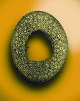
A Micro-Drill Tool Tip is another image turned into art and was taken using the ZEISS LSM 700 confocal microscope. This confocal laser scanning microscope has a compact design and performs fluorescence measurements, creates optical sections of samples, and combines them into 3D image stacks. ZEN imaging software shortens the training period and stores the settings for all users.
An image of the Birefringence of Glucose Monohydrate was taken with the ZEISS Axiovert.A1 200M This model has been replaced with the ZEISS Axio Observer, an inverted microscope with extensive performance capabilities. Highly configurable, this microscope has a variety of excitation and detector options and can be upgraded with high speed, confocal and superresolution. The ZEISS Axio Observer is equipped with environmental controls, for long term, gentle imaging of living samples.
The final piece of art was created using the ZEISS LSM 710 Laser Scanning Confocal Microscope. IGB researchers used it to take an image of a 3D cleared and tiled Miscanthus. The prerequisite for every demanding application in laser scanning microscopy is enhanced sensitivity and reduced background noise. The ZEISS LSM 710 suppresses noise from excitation laser light to deliver class leading signal to noise, even with tricky preparations, such as those with dense 3D tissue or cells growing directly on metallic substrates.
About the Institute for Genomic Biology
The Institute for Genomic Biology (IGB) at the University of Illinois is dedicated to researching significant problems facing humanity. Treating chronic human diseases, managing new and emerging pests and pathogens, and maintaining abundant and healthy food and water supplies are just a few of the big challenges being addressed at IGB. Established in 2003, the Institute for Genomic Biology harnesses new and emerging genomics technologies to advance basic biology research and to apply those innovations to create useful products and high-tech jobs. Learn more at www.igb.illinois.edu or contact: Melissa McKillip, (217) 333-4619, mmckilli@illinois.edu.
ZEISS
The Carl Zeiss Group is an international leader in the fields of optics and optoelectronics. In fiscal year 2011/12 the company's approximately 24,000 employees generated revenue of nearly 4.2 billion euros. In the markets for Industrial Solutions, Research Solutions, Medical Technology and Consumer Optics, ZEISS has contributed to technological progress for more than 160 years and enhances the quality of life of many people around the globe. The Carl Zeiss Group develops and produces planetariums, eyeglass lenses, camera and cine lenses and binoculars as well as solutions for biomedical research, medical technology and the semiconductor, automotive and mechanical engineering industries. ZEISS is present in over 40 countries around the globe with about 40 production facilities, over 50 sales and services locations and approximately 20 research and development sites. Carl Zeiss AG is fully owned by the Carl Zeiss Stiftung (Carl Zeiss Foundation). Founded in 1846 in Jena, the company is headquartered in Oberkochen, Germany.
Microscopy
The Microscopy business group at ZEISS is the world's only manufacturer of light, X-ray and electron microscopes. The company's extensive portfolio enables research and routine applications in the life and materials sciences. The product range includes light and laser scanning microscopes, X-ray microscopes, electron and ion microscopes and spectrometer modules. Users are supported for software for system control, image capture and editing. The Microscopy business group has sales companies in 33 countries. Application and service specialists support customers around the globe in demo centers and on site. The business group is headquartered in Jena, Germany. Additional production and development sites are in Oberkochen, Göttingen and Munich, as well as in Cambridge in the UK and Peabody, MA and Pleasanton, CA in the USA. The company has around 2,800 employees and generates revenue of 650 million euros.
Attention publishers: Please forward all Sales Inquiries to: Maya Everett, Carl Zeiss Microscopy, (914) 681-7782, maya.everett@zeiss.com