A Complete Concept in Personalisation
Introducing the new Olympus cellSens life science imaging software
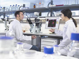
Hamburg, - Olympus has today introduced the new cellSens software for life science imaging applications. Consisting of three different packages: cellSens Entry, cellSens Standard and cellSens Dimension, all user requirements can be met with ease. Incorporating a unique user-definable interface, the imaging process can be personalised from start to finish, for complete control over the entire experiment. As a result, only the required tools are present at each stage, enabling everyone to image with ease, regardless of experience levels. Furthermore, each package integrates seamlessly with Microsoft Windows and Office programmes, making the advanced functionality of the Olympus cellSens family extremely user-friendly.
cellSens Entry
With the ability to perform simple image acquisition and documentation, cellSens Entry is ideal for basic image capture. The unique customisable interface provides complete control over a range of Olympus digital cameras to obtain live images in a number of formats, including AVI movies. Users can therefore capture the image they require, with ease. The cellSens Entry package can perform straight-forward post-capture processing such as light intensity and white balance adjustments, and its layers file format enables the addition of arrows and annotation text without affecting the original image channels.
cellSens Standard
Building on the concept of cellSens Entry, the cellSens Standard software enables more advanced image capture, including time-lapse TWAIN acquisition. With advanced hardware control, processes such as objective and filter changes, focusing and internal shutter control can all be motorised, enhancing the user's ability to precisely manage the acquisition. Supplied with extended geometry functions, images are easily manipulated via mirroring, rotation, resizing, cropping and channel shifting. Creating additional flexibility, cellSens Standard can also convert bit-depth and colour-space settings to meet the capabilities of the computer system as well as the requirements of the application. Further image processing tools are available for contrast adjustment, smoothing (lowpass), image sharpening, as well as noise and shading correction. Increasing this versatility, the software can also process multi-dimensional images for extremely accurate depiction of sections through a tissue by manipulating channels as well as combining or extracting specific frames. Within the images, interactive measurement functions can be executed on a separate image layer. Extracted numerical values such as size, distance or area can subsequently be exported to Microsoft Excel for in-depth statistical analysis.
cellSens Dimensions
Designed to offer the control and processing requirements for microscope-based experimental systems, the cellSens Dimensions software provides a broad range of advanced features as well as specialised, optional solution modules. It is possible to successfully conduct a range of complex and highly sophisticated experiments, from extended focal imaging to multiple image alignment and multi-position imaging. In addition, live images can be transferred directly to the web using the netcam functionality, enabling rapid discussion with colleagues, wherever they are in the world. With the ability to control a wide range of Olympus and non-Olympus hardware, , advanced and precise time-lapse experiments can be conducted. On the resulting time stacks, kinetic analysis and threshold based object analysis are possible. Once data has been obtained, the unique report composer uses Word templates to generate user-defined reports that retrieve images and data directly from the cellSens database.
Solution modules
A series of optional modules enables the user to further expand functionality. For example, the multichannel 5D solution enables the automatic acquisition of images in five dimensions: X, Y, Z, multi-channel fluorescence with transmission overlay, time-lapse and multi-position. Other modules include: the multi-position solution for automated panorama imaging functionality, the database solution to add a client-server database; the deconvolution solution to remove out-of-focus blur for sharp image definition; and the object detection solution for precise threshold-based object detection as well as spectral un-mixing of brightfield images.
Please contact:
OLYMPUS EUROPA HOLDING GMBH
Dr Friederike Lehmann
Marketing Communications
Tel: +49 40 2 37 73 - 5406
Fax: +49 40 2 37 73 - 4647
E-mail: microscopy@olympus-europa.com
www.microscopy.olympus.eu